Simplifying common cardiology investigations
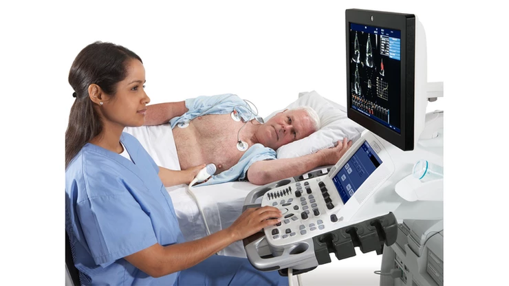
Q. What is 2 D Echocardiography and Colour Doppler?
A. 2 D Echocardiography is a non invasive test to visualise the heart. It works on the principle of Sonar. It sends sound waves(outside the audible frequency) through a transducer. The same sound waves are reflected inside the chest by the heart and other structures, and are received back by the transducer. The computer calculates the delay between the sent and the received sound waves, and constructs a picture of the heart in real time.
Q. What can be seen by a 2 D Echocardiography and Colour Doppler?
A. 2 D Echocardiography can be used to visualise the following:
1. The contractility and "pumping power" of the heart. Patients who have had a heart disease may have decreased pumping power of the heart, either localised, or generalised.
2. Valves of the heart: Valve structure, Valve narrowing, or valve leak can be identified.
3. Fluid around the heart (Pericardial Effusion) can be identified.
4. "Holes" between cardiac chambers, and abnormal communication of blood between chambers is seen. These can be then corrected,
5. High pressure in lung arteries, due to respiratory diseases or heart diseases can be diagnosed.
6. Abnormailities of big blood vessels like Aorta can be detected.
7. Tumors or blood clots inside the heart can be detected.
Apart from this, chamber sizes can be measured. Wall thickness can be measured, and several sophisticated calculations can be performed.
This test has no side effects and is 100% safe. It can be done in 10-15 minutes.
SoBo Heart Clinic has a high end , Cardiology specific 2 D Echocardiography machine from GE, which gives fantastic resolution